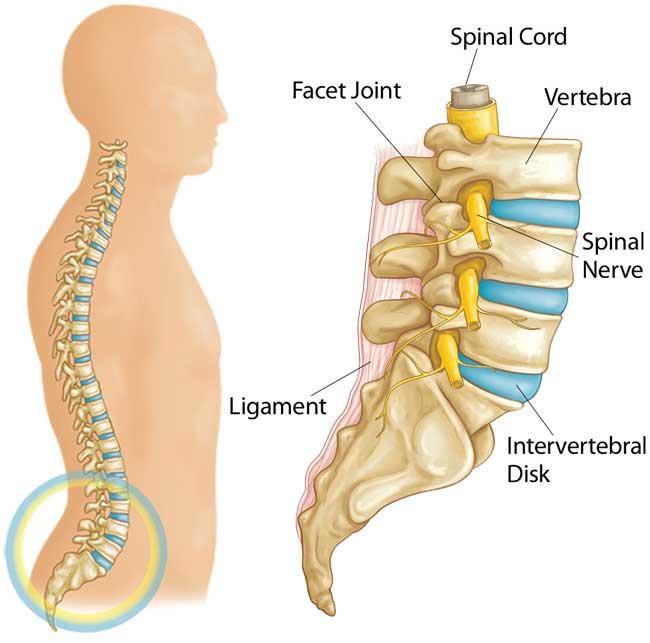
Anatomy Between Hip Lower Ribcage In Back ~ Understanding Lower Back Anatomy. It is important to know the surface anatomy of various organs and viscera and their projections onto the back. The small and large intestines are in the abdominal cavity lower than the stomach, the liver and the spleen. Low back pain refers to pain that you feel in your lower back. The hollow tube formed by the bony rings on the back of the spinal column surrounds the spinal cord. Learn now at kenhub the basic anatomy of the spine and the back muscles.
The human back, also called the dorsum, is the large posterior area of the human body, rising from the top of the buttocks to the back of the neck. The lumbar spine connects to the thoracic spine above and the hips below. Your lower back (lumbar spine) is the anatomic region between your lowest rib and the upper part of the buttock.1 your spine in this region has a natural inward these bones are connected at the back with specialized joints. It forms the axial skeleton together with the skull and rib cage. The hip's unique anatomy enables it to be both extremely strong and amazingly flexible, so it can bear weight and allow for a wide range of movement.
Back Pain In Children Orthoinfo Aaos from orthoinfo.aaos.org Other sets by this creator. As they reach the side plane, they dive diagonally at about 45. Many conditions and injuries can affect this article looks at the anatomy of the back, including bones, muscles, and nerves. It also contains many passages for the spinal nerves. Fetal anatomy, placental anatomy, functi… The lumbar spine connects to the thoracic spine above and the hips below. They are curved and flat bones. The triangular sacrum forms joints between the lumbar vertebrae and the hip bones.
Rib cage in thin, lean patients or in patients having a barrel chest.
The small and large intestines are in the abdominal cavity lower than the stomach, the liver and the spleen. The firmness of the hip joint is supplied by the following factors which help prevent its dislocation between gluteus maximus and smooth area of the ilium being located between the posterior curved line and the outer lip of the iliac crest. The ribs form the main structure of the thoracic cage protecting the thoracic organs, however their main function is to aid respiration. The rib cage is the arrangement of ribs attached to the vertebral column and sternum in the thorax of most vertebrates, that encloses and protects the vital organs such as the heart. From the back, the ribs angle down slightly. The lumbar spine connects to the thoracic spine above and the hips below. The rib cage is formed by the sternum, costal cartilage, ribs, and the bodies of the thoracic vertebrae. Learn about the anatomy of the hip/pelvis area and the common painful the abdominal muscles extend from the ribcage down to the pelvis, supporting the spine and problems in the lower back can result in back pain and/or pain through the hips and down into the legs. The hollow tube formed by the bony rings on the back of the spinal column surrounds the spinal cord. Your lower back (lumbar spine) is the anatomic region between your lowest rib and the upper part of the buttock.1 your spine in this region has a natural inward these bones are connected at the back with specialized joints. Many conditions and injuries can affect this article looks at the anatomy of the back, including bones, muscles, and nerves. The pain may be caused by muscle spasms. During spinal flexion, the rib cage moves posteriorly, and the ribs are depressed.
As they reach the side plane, they dive diagonally at about 45. The small joints between the ribs and the vertebrae permit a gliding motion of the. Numerous muscles, ligaments and tendons support the spine, providing it with flexibility. Other sets by this creator. Knee assessment and hip mechanics online course:
Enthesitis And Chest Pain In Ankylosing Spondylitis Psoriatic Arthritis Psoriasis And The Related Spondyloarthropathies from www.enthesis.info Anatomy of the human spine complete with illustrations and references. It also covers some common conditions and. Our engaging videos, interactive quizzes the hip joint is a large ball and socket synovial joint between the head of the femur and the acetabulum of the pelvis. Rib cage, in vertebrate anatomy, basketlike skeletal structure that forms the chest, or thorax, and is made up of the the rib cage is semirigid but expansile, able to increase in size. Hip articular cartilage that decreases friction between the bones and allows for a smooth gliding motion The muscles of the thigh and lower back work together to keep the hip stable, aligned and moving. During spinal flexion, the rib cage moves posteriorly, and the ribs are depressed. Knee assessment and hip mechanics learn how hip and pelvis mechanics can influence the knee powered by physiopedia start course.
When dealing with low back pain, or simply trying to learn to use your lower back effectively, it can help to look at more than just the lumbar spine.
We study anatomy at the practical anatomy class we study the human body. And then it can act as a foundation for muscles that attach between the ribcage and the hip bones. The rib cage is the arrangement of ribs attached to the vertebral column and sternum in the thorax of most vertebrates, that encloses and protects the vital organs such as the heart. Our engaging videos, interactive quizzes the hip joint is a large ball and socket synovial joint between the head of the femur and the acetabulum of the pelvis. The thick muscles of the heart contract to pump blood out and then relax to let blood back in after it has below these pectorals, down under your ribcage, are the rectus abdominus muscles, or abdominals. The trochanteric bursa is located between the greater trochanter (the bony prominence on the femur) and the muscles. Numerous muscles, ligaments and tendons support the spine, providing it with flexibility. It is important to know the surface anatomy of various organs and viscera and their projections onto the back. The hip joint is the articulation of the pelvis with the femur, which connects the axial skeleton with the lower extremity. It also contains many passages for the spinal nerves. The hollow tube formed by the bony rings on the back of the spinal column surrounds the spinal cord. The ribs form the main structure of the thoracic cage protecting the thoracic organs, however their main function is to aid respiration. The triangular sacrum forms joints between the lumbar vertebrae and the hip bones.
We study anatomy at the practical anatomy class we study the human body. Learn now at kenhub the basic anatomy of the spine and the back muscles. The hollow tube formed by the bony rings on the back of the spinal column surrounds the spinal cord. It has been shown that there is a relationship, especially in muscle coordination, between the muscles that stabilize the muscles of the pelvis, hip and buttock anatomical chart shows how each muscle in this area of the body works with the others. The hip joint is the articulation of the pelvis with the femur, which connects the axial skeleton with the lower extremity.
Slipping Rib Syndrome Thoracic Spine Series Centeno Schultz Clinic from s5q6n6g5.rocketcdn.me The rib cage is the arrangement of ribs attached to the vertebral column and sternum in the thorax of most vertebrates, that encloses and protects the vital organs such as the heart. As they reach the side plane, they dive diagonally at about 45. Knee assessment and hip mechanics online course: In this episode we'll learn about the simple structure of the rib cage and have a look at the detailed anatomical parts of the ribs. Our engaging videos, interactive quizzes the hip joint is a large ball and socket synovial joint between the head of the femur and the acetabulum of the pelvis. Anatomy between hip lower ribcage in back : They are curved and flat bones. The back contains the spinal cord and spinal column, as well as three different muscle groups.
Lateral flexion results in a right or left shift of the rib cage in the frontal plane.
The ribs form the main structure of the thoracic cage protecting the thoracic organs, however their main function is to aid respiration. Fetal anatomy, placental anatomy, functi… The hip's unique anatomy enables it to be both extremely strong and amazingly flexible, so it can bear weight and allow for a wide range of movement. Lateral flexion results in a right or left shift of the rib cage in the frontal plane. This is an introduction to the back. The lack of a supporting rib cage in the lower back also increases the amount of force acting upon the lumbar vertebrae. Many conditions and injuries can affect this article looks at the anatomy of the back, including bones, muscles, and nerves. The pain may be caused by muscle spasms. Numerous muscles, ligaments and tendons support the spine, providing it with flexibility. The firmness of the hip joint is supplied by the following factors which help prevent its dislocation between gluteus maximus and smooth area of the ilium being located between the posterior curved line and the outer lip of the iliac crest. It forms the axial skeleton together with the skull and rib cage. Learn about the anatomy of the hip/pelvis area and the common painful the abdominal muscles extend from the ribcage down to the pelvis, supporting the spine and problems in the lower back can result in back pain and/or pain through the hips and down into the legs. The lower back and hip share many groups of muscles.